What do you understand about PDB or
also known as Protein Data Bank ?
also known as Protein Data Bank ?
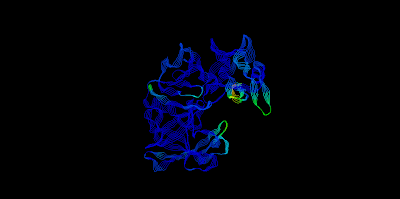
- The Protein Data Bank (PDB) is a repository for the 3-D structural data of large biological molecules, such as proteins and nucleic acids.
- The data, typically obtained by X-ray crystallography or NMR spectroscopy and submitted by biologists and biochemists from around the world, are freely accessible on the Internet via the websites of its member organisations (PDBe,[1] PDBj,[2] and RCSB[3]).
- The PDB is overseen by an organization called the Worldwide Protein Data Bank, wwPDB.
Types of Protein :
- LexA
- Repressor LexA or LexA is a repressor enzymethat represses SOS response genes coding for DNA polymerases required for repairing DNA damage. LexA is intimately linked to RecA in the biochemical cycle of DNA damage and repair. RecA binds to DNA-bound LexA causing LexA to cleave itself in a process called autoproteolysis.
- Any further information about LexA, you may click this : http://www.rcsb.org/pdb/explore/explore.do?structureId=1JHF
2. Trypsin
- Trypsinis a serine protease found in the digestive system of many vertebrates, where it hydrolyses proteins. Trypsin is produced in the pancreas as the inactive proenzyme trypsinogen. Trypsin cleaves peptide chains mainly at the carboxyl side of the amino acids lysine or arginine, except when either is followed by proline.
- It is used for numerous biotechnological processes. The process is commonly referred to as trypsin proteolysis or trypsinisation, and proteins that have been digested/treated with trypsin are said to have been trypsinized.
- Any further information about Trypsin, you may click this : http://www.rcsb.org/pdb/explore/explore.do?structureId=2PTC
- High-temperature requirement A (HtrA), a highly conserved family of serine protease, plays crucial roles in protein quality control in prokaryotes and eukaryotes.The HtrA protein contains a C-terminal PDZ domain that mediates the proteolytic activity.
- The structure of HtrA PDZ domain, which contains three α-helices and five β-strands, illustrates conservation within the canonical PDZ domains.
- The ligand binding surface and the critical residues responsible for ligand binding of HtrA PDZ domain by chemical shift perturbation and site-directed mutagenesis.
- Any further information about HtrA, you may click this : http://www.rcsb.org/pdb/explore/explore.do?structureId=2L97
4. Pepsin
- Pepsin (from the Greek πέψη, pepsi, meaning digestion) is an enzyme whose zymogen (pepsinogen) is released by the chief cells in the stomach and that degrades food proteins into peptides.
- Pepsin is one of three principal protein-degrading, or proteolytic, enzymes in the digestive system, the other two being chymotrypsin and trypsin. The three enzymes were among the first to be isolated in crystalline form.
- During the process of digestion, these enzymes, each of which is specialized in severing links between particular types of amino acids, collaborate to break down dietary proteins into their components, i.e., peptides and amino acids, which can be readily absorbed by the intestinal lining. Pepsin is most efficient in cleaving peptide bonds between hydrophobic and preferably aromatic amino acids such as phenylalanine, tryptophan, and tyrosine
- Any further information about Pepsin, you may click this : http://www.rcsb.org/pdb/explore/explore.do?structureId=3UTL
- Amylaseis an enzyme that catalyses the breakdown of starch into sugars. Amylase is present in the saliva of humans and some other mammals, where it begins the chemical process of digestion. Foods that contain much starch but little sugar, such as rice and potato, taste slightly sweet as they are chewed because amylase turns some of their starch into sugar in the mouth.
- The pancreas also makes amylase (alpha amylase) to hydrolyse dietary starch into disaccharides and trisaccharides which are converted by other enzymes to glucose to supply the body with energy.
- Plants and some bacteria also produce amylase. As diastase, amylase was the first enzyme to be discovered and isolated (by Anselme Payen in 1833).[1] Specific amylase proteins are designated by different Greek letters. All amylases are glycoside hydrolases and act on α-1,4-glycosidic bonds.
- Amylase can be divided into 3 types which are : -
| Classification | Description | |
|---|---|---|
| α-Amylase | The α-amylases are calcium metalloenzymes, completely unable to function in the absence of calcium. By acting at random locations along the starch chain, α-amylase breaks down long-chain carbohydrates, ultimately yielding maltotriose and maltose from amylose, or maltose, glucose and "limit dextrin" from amylopectin. Because it can act anywhere on the substrate, α-amylase tends to be faster-acting than β-amylase. In animals, it is a major digestive enzyme, and its optimum pH is 6.7-7.0 | |
| β-Amylase | β-amylase is also synthesized by bacteria, fungi, and plants. Working from the non-reducing end, β-amylase catalyzes the hydrolysis of the second α-1,4 glycosidic bond, cleaving off two glucose units (maltose) at a time. During the ripening of fruit, β-amylase breaks starch into maltose, resulting in the sweet flavor of ripe fruit. β-amylase is present in an inactive form prior to germination, whereas α-amylase and proteases appear once germination has begun The optimum pH for β-amylase is 4.0-5.0 | |
| γ-Amylase | γ-Amylase will cleave α (1 - 6) glycosidic linkages, as well as the last α (1 - 4) glycosidic linkages at the non recuding end of amylose and amylopectin, yielding glucose. The γ-Amylase has most acidic pH optimum because it is most active around pH 3. |
- Any further information about Amylase, you may click this : http://www.rcsb.org/pdb/explore/explore.do?structureId=1BAG


No comments:
Post a Comment